
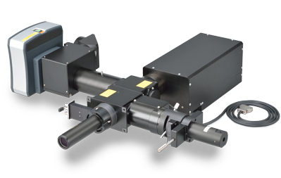
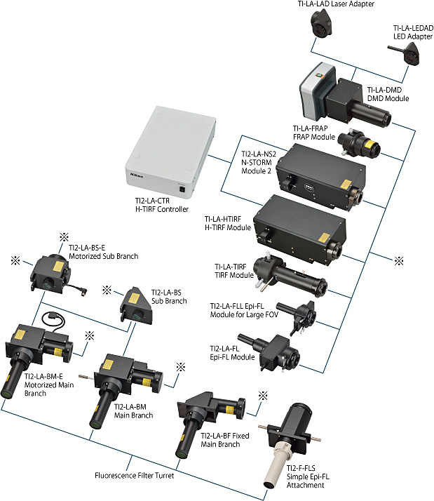
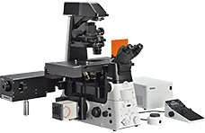
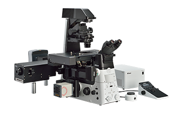
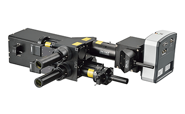
模块化照明系统具有极高的灵活性和可扩展性
尼康Ti2-LAPP系统为全内反射荧光(TIRF),光活化/转换,光漂白和落射荧光以及超分辨显微镜(N-STORM)提供模块化照明。 每个模块可以灵活组合,构建针对个别研究需求进行优化的显微镜系统。 例如,可以将多个TIRF模块并入到单个显微镜中用于各向异性实验和快速,多角度TIRF成像。结合Ti2的多层结构,多达五个照明模块可以并入单个显微镜(例如,两个TIRF,FRAP,DMD和落射荧光模块都可以集成到一个Ti2中)。
Description
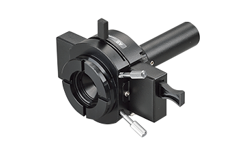
DMD模块
实现同时多点光活化
DMD模块可实现用户指定的图案和位置的光激活和光电转换,而传统的FRAP单元仅能够对单个手动定位的光点进行光活化。 DMD照明形状,尺寸,位置和数量可以使用NIS-Elements软件自由定制。该功能允许研究人员在单个细胞或多个细胞中光学标记细胞或蛋白质群体的子集,以追踪其行为。DMD模块也最适合于光遗传学实验,其中高度定制的ROI可用于光学诱导细胞或蛋白质群体或亚群的功能变化。DMD模块可用于激光照明或较少光毒性的LED照明。
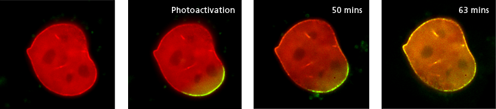
A mouse embryonic fibroblast co-expressing mCherry-tagged lamin A (red) and photo-activatable GFP-tagged lamin A was photo-converted (green) in the lower right region using the DMD module and 405 nm LED light. Time-lapse images were captured using the epi-fluorescence illuminator. By photoactivating a sub-population of the lamin proteins, one can observe their dynamics and subunit-exchange behavior.
Image courtesy of Drs. Takeshi Shimi and Bob Goldman, Northwestern University Medical School
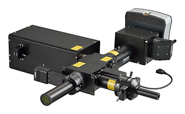
H-TIRF模块
现在可以进行全自动TIRF调整和观察
激光对于TIRF观察的理想入射角和焦点取决于样本和观察条件。调整入射角度并聚焦以实现TIRF需要技能和经验。H-TIRF模块通过监测反射光束自动调整激光的聚焦和入射角,用于TIRF观察。通过NIS-Elements软件中的自动对准功能进行自动激光聚焦调整和入射角度调整。可以保存和再现渐逝场的入射角和穿透深度,用于后续实验,以确保一致的成像结果。
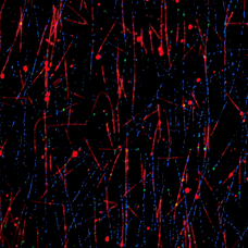
An in vitro preparation of fluorescently-labeled microtubules (tetramethylrhodamine and Alexa 647) and tubulin binding proteins (Alexa 488) was imaged in three different wavelengths using the H-TIRF illuminator and the gradation ND filter. Incident angles can be automatically adjusted for multiple wavelengths.
The video of this image is in the “Sample Images” page.
Image courtesy of Melissa Hendershott and Dr. Ron Vale, University of California, San Francisco
没有灰度级ND滤镜,TIRF照明在FOV中显示高斯分布,中心是最亮的。
使用灰度级ND滤镜,实现了非常均匀的TIRF照明。
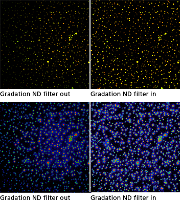
An in vitro preparation of a bi-lipid membrane containing Alexa 488 (green)- and Alexa 561 (red)-tagged membrane-associated proteins was imaged using the H-TIRF illuminator and two different wavelengths (dual color TIRF). The proteins aggregate to form clusters that are visualized as circular structures (the top row).
The lower row shows the 488 channels displayed using a rainbow look-up table where different intensities are represented as different colors.
The H-TIRF illuminator can be used to automatically achieve optimal incident angles for the different wavelengths.
Image courtesy of Drs. Xiaolei Su and Ron Vale, University of California, San Francisco
N-STORM模块
实现比常规光学显微镜提高10倍的分辨率
配备照明场(1x,2x,4x)电动开关以及自动对准功能,该模块可与Ti2-LAPP系统进行N-STORM超分辨率成像。这提供了大约20nm的令人难以置信的图像分辨率,其是常规光学显微镜的极限的10倍或更大。利用STochastic光学分辨显微镜(STORM),可以在分子水平上深入了解蛋白质 – 蛋白质的相互作用。
FRAP模块
用于分析细胞内-蛋白质动力学
使用这种FRAP模块,可以进行光漂白和光激活/转换实验以及使用高帧率,高灵敏度的相机进行检测。该模块可以点亮细胞中的目标位置,为研究细胞内蛋白质动力学而不使用点扫描共焦显微镜提供了低成本高效益的方法。
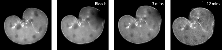
A mouse embryonic fibroblast expressing mCherry-lamin A was spot-photobleached in the upper right corner of the nucleus using the FRAP module to study the dynamics of lamin A molecules. Time-lapse images were acquired using the epi-fluorescence illuminator.
Image courtesy of Drs. Takeshi Shimi and Bob Goldman, Northwestern University Medical School
TIRF模块
用于观察细胞膜动力学和单分子
手动TIRF模块包括一个灰度级ND滤镜(类似于H-TIRF模块),能够在整个视野内实现均匀的TIRF照明。使用高灵敏度摄像机,可以使用该TIRF照明器在细胞膜内和附近成像单分子和蛋白质的动力学。
大视野落射荧光模块
适用于带有大型传感器的相机的荧光成像
用于大型FOV的EPI FL模块是Ti2-LAPP系统的基本落射荧光照明器,专为使用Ti2倒置显微镜的大型FOV成像应用而设计。 即使它具有紧凑的设计,它在落射光观察期间提供无与伦比的大的25mm视野。它配备了石英复眼镜片,以确保整个视野的均匀强度,并在广泛的光谱(包括紫外线)下提供高透射率。
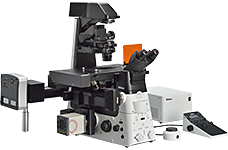
灵活组合模块
Ti2-LAPP系统的模块化和灵活配置功能为个人研究需求提供定制成像解决方案。模块也可以轻松交换或添加,以适应不断变化的实验需求,这是实验室的重要功能,具有不断发展的研究方向和多用户核心设施。例如,通过在单TIRF配置中添加第二个TIRF模块,用户可以轻松执行各向异性实验和快速,多角度TIRF实验。添加诸如DMD或FRAP模块之类的光激活/转换模块可以跟踪蛋白质群体的亚分支,从而提供对成像整个群体时的虚假信息的蛋白质行为的见解。
双层配置功能
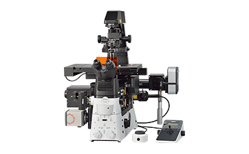
利用尼康Ti2的层次结构,模块可以作为两层独立的层叠,每层有多个模块。使用双层配置可为每个照明模块实现最佳的过滤器配置。例如,通过将H-TIRF模块放置在下层中,并且在上层中设置DMD模块,可以将特定于TIRF成像和光活化的单独的滤光块同时用于它们各自位于下层和上层的转轮中。该配置可实现最佳的滤光块选择,并提高实验精度,同时保持最高的采集速度。
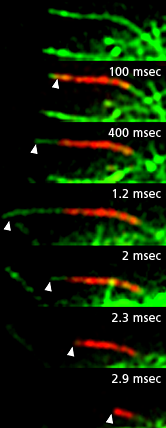 |
A Drosophila S2 cell expressing EOS-tubulin. The end of a single microtubule was photoconverted using the DMD module and 405 nm LED light. Time-lapse images in dual color TIRF were acquired using the H-TIRF illuminator. The addition of unconverted, green tubulin to the growing end of the photoconverted red microtubule, and shortening (and eventual disappearance) of the photoconverted segment demonstrate the dynamic instability property of microtubules. Arrowheads mark the growing and shrinking end of the photoconverted microtubule. |
|---|