
令人惊叹的深度 – A1 MP + / A1R MP +可以显著地观察生物体内的超深度动力学。
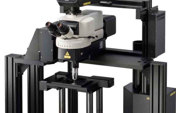
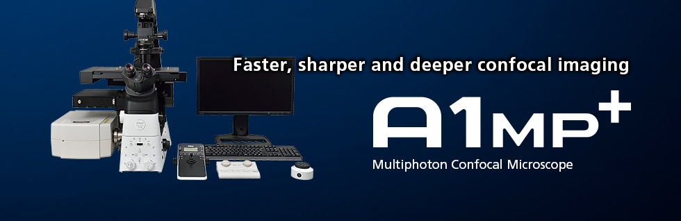
A1 MP+ / A1R MP+多光子共聚焦显微镜从生物体内更深层次地提供更快更清晰的成像,延伸了生物科学中传统研究技术的界限。它们与直立和倒置显微镜兼容,并为大脑研究,其他神经科学应用和活体标本的体内成像提供最佳的多光子成像配置。
Description
超高灵敏度GaAsP NDD的活体内深度成像
GaAsP NDD配备GaAsP PMT,具有比多碱PMT更高的信噪比和更高的灵敏度,并且可以对较深的活体样品进行清晰的成像。获得高信噪比图像的能力可以实现更快的成像和更高质量的Z-stack成像。其高灵敏度允许以较少的激光功率采集荧光信号,导致对活体样本的光损伤较少。
尼康A1 MP+和A1R MP+可配置1080nm波长或1300nm波长,可实现高达1.4mm的深度成像。
NDD尽可能靠近样本,以便检测来自深层活体标本内的最大散射信号。通过检测反射和透射的荧光信号,用于Ni-E/FN1立式显微镜的表观和双光子GaAsP NDD的组合可以获得明亮和高信噪比的图像。
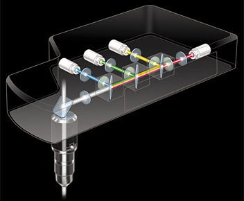
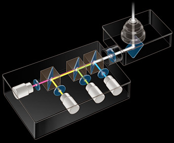
Left:4通道反射 GaAsp NDD;Right:4通道透射GaAsp NDD
兼容1300nm波长的GaAsp NDD进行活体小鼠脑深度成像
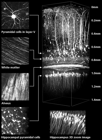
In vivo imaging of an anesthetized YFP-H mouse (4-week-old) via open skull method. Visualization of the entire layer V pyramidal neurons and the deeper hippocampal neurons. Deep imaging achieved for 3-dimensional imaging of hippocampal dendrites up to 1.4 mm into the brain.
Captured with episcopic GaAsP NDD for 1300 nm and CFI75 Apochromat 25XC W 1300 objective (NA 1.10, WD 2.0 mm)
Excitation wavelength: 1040 nm
Photos courtesy of: Drs. Ryosuke Kawakami, Terumasa Hibi and Tomomi Nemoto, Research Institute for Electronic Science, Hokkaido University
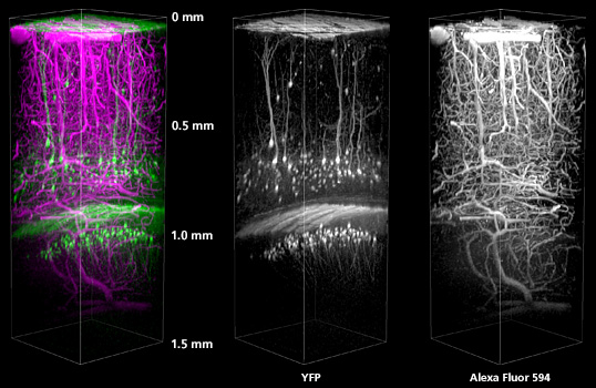
The cerebral cortex of an anesthetized YFP-H mouse (4-week-old) was studied with the open skull method.
Alexa Fluor 594 was injected into the tail vein to visualize the blood vessel.
Excitation wavelength: 1100 nm
Objective: CFI75 Apochromat 25XC W 1300 (NA 1.10, WD 2.0)
Photos courtesy of: Drs. Ryosuke Kawakami, Terumasa Hibi and Tomomi Nemoto, Research Institute for Electronic Science, Hokkaido University
双波长红外激光同时激发成像
A1 MP+和A1R MP+可用于兼容双波长红外激光同时激发的系统。
将该系统与具有双波长同时输出(700-1300nm的主可调输出和1040nm的辅助固定输出)的飞秒IR脉冲激光器结合,能够在活细胞内的深部区域中同时激发和成像两种不同的染料。
斑马鱼的两个波长同时激发成像
Three dimension images of 1 dpf zebrafish transgenic line, Tg[h2afv:GFP; EF1α: mCherry-zGem]. After breeding under the treatment of Phenyltiourea (PTU), which inhibits melanin synthesis, whole body was clarified with optical clearing solution LUCID-A. This transgenic line visualizes proliferating cells and chromatin with mCherry (red) and GFP (green), respectively.
Excitation wavelength: 900 nm and 1040 nm
Objective: CFI75 Apochromat 25XC W 1300 (NA 1.10, WD 2.0)
Photos courtesy of: Drs. Toshiaki Mochizuki and Ichiro Masai, Developmental Neurobiology Unit, Okinawa Institute of Science and Technology Graduate University
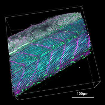
Lateral view of trunk of zebrafish transgenic line Tg[h2afv:GFP; EF1α: mCherry-CAAX] at 34 hpf. After breeding under the treatment of Phenyltiourea (PTU), which inhibits melanin synthesis, whole body was clarified with optical clearing solution LUCID-A. This transgenic line visualizes cell membrane and chromatin with mCherry (purple) and GFP (green), respectively. SHG (blue) indicates muscle fibers.
Excitation wavelength: 900 nm for SHG, GFP and 1040 nm for mCherry
Objective: CFI75 Apochromat 25XC W 1300 (NA 1.10, WD 2.0)
Photos courtesy of: Drs. Toshiaki Mochizuki and Ichiro Masai, Developmental Neurobiology Unit, Okinawa Institute of Science and Technology Graduate University
可选择的扫描头启用高速,高品质的成像
A1R MP+是混合扫描头,它兼有高分辨率检流计(非共振)扫描器和超高速共振扫描器。 其混合扫描头允许以显示细胞动力学和相互作用所需的超快速度的成像和光活化。 A1 MP+配备了检流计扫描器,用于高分辨率成像。 A1R MP+和A1 MP+都可配置为1080nm和1300nm的两个波长。
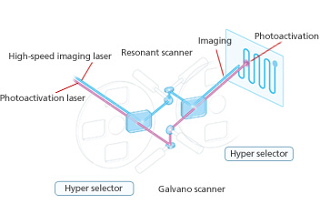
A1R MP+ 混合扫描头
超高速成像高达每秒420帧
A1R MP+的共振扫描器具有7.8kHz的超高频共振频率,可实现高达420 fps(512 x 32像素)的快速成像。尼康的光学像素时钟发生系统即使在高速下也能保证稳定,几何正确和均匀的照明成像。 这使得能够成功地观察体内快速变化,例如活体生物中的反应,动力学和细胞相互作用。
此外,高分辨率共振扫描器提供1024 x 1024像素(15 fps)的高分辨率成像。
| 1D 扫描 | 15,600 lps |
|---|---|
| 2D scanning | 420 fps (512 x 32 pixels) |
| Full frame scanning | 60 fps (256 x 256 pixels) 30 fps (512 x 512 pixels) 15 fps (1024 x 1024 pixels) |
可视化活体微循环
Blood cells in blood vessels within a living organism were excited by a femtosecond pulsed IR laser with the A1R MP+’s ultrahigh-speed resonant scanner, and their movements were simultaneously captured in three successive fluorescence images at 30 fps (30 msec), with three separate color channels.
Three fluorescent probes are simultaneously excited and imaged–nucleus (blue), endothelium (green), and plasma (red). The long-wavelength ultrafast laser in combination with the ultrahigh-speed resonant scanner effectively reduces photodamage and makes time resolved multiphoton imaging of biomolecules possible.
Image resolution: 512 x 512 pixels, Image acquisition speed: 30 fps, Objective: water immersion objective 60X
Photos courtesy of: Dr. Satoshi Nishimura, Center for Molecular Medicine, Jichi Medical University
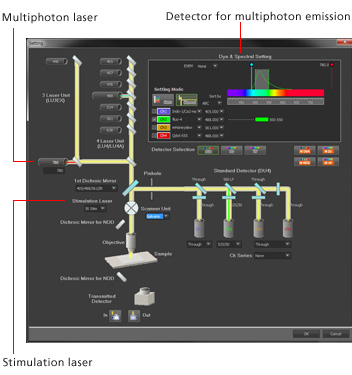
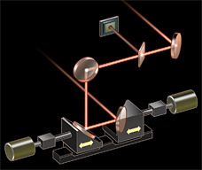
改变多光子激发波长时的自动激光对准
当多光子激光波长或组速度色散预补偿改变时,位于物镜后孔的位置指向的多光子激光束也可能改变,导致图像上的不均匀强度或IR和可见激光光路之间的微小的未对准。
传统上,验证IR激光束指向和设置对准一直是困难的。 Nikon的A1 MP+ / A1R MP+系列自动激光对准功能,容纳在多光子激发光路的入射光学单元中,可以通过NIS-Elements C中的单次点击自动最大化IR激光对准。
(在800 nm – 1300 nm波长范围内可进行自动激光对准)
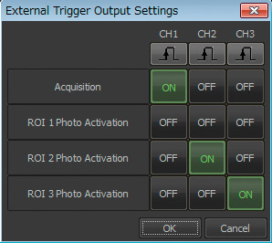
统一采集分析软件平台
尼康的统一软件平台,NIS-Elements为共聚焦成像提供了直观的工作流程。结合JOBS和照明序列等图形编程工具,可以根据任何应用需求,全面定制综合操作环境。